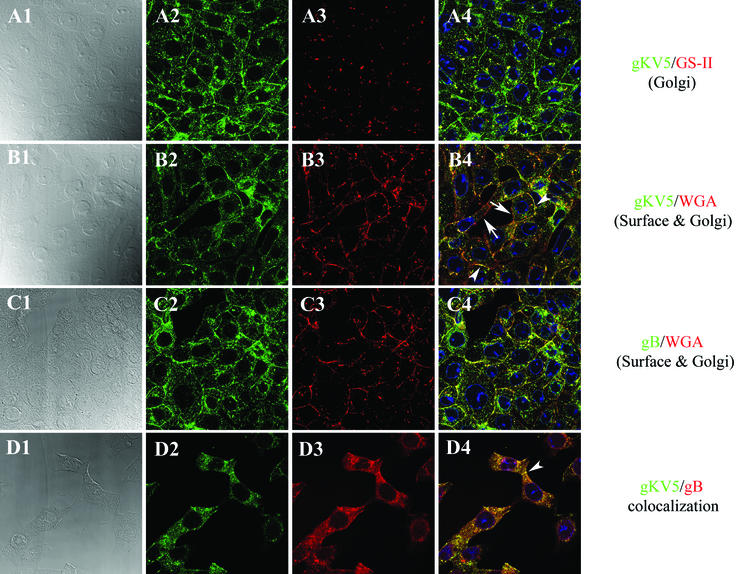
FIG. 4.
Subcellular distribution of gK and gB in gKV5DIII virus-infected cells. Vero cells were infected with gKV5DIII at an MOI of 5. Cells were fixed and processed for confocal microscopy; panels show staining with anti-V5 (green: A2, A4, B2, B4, D2, and D4), anti-gB (green: C2 and C4), or anti-gB (red: D3 and D4) antibodies at 12 hpi. Cellular organelles were counterstained with TO-PRO3 to specifically stain the nucleus (blue: A4, B4, C4, and D4) and with either lectin GS-II, which specifically stains the Golgi apparatus (red: A3 and A4), or lectin WGA to label Golgi and plasma membranes (red: B3, B4, C3, and C4). Corresponding DIC images of cells are as shown (A1, B1, C1, and D1). Superimpositions of red and green images for each group of images are shown (A4, B4, C4, and D4).

