FIG. 5.
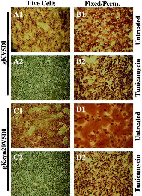
Cell surface immunohistochemical detection of gK in gKV5DI- or gKsyn20V5DI-infected cells. Vero cells were infected with gKV5DI (A1, B1, A2, and B2) or gKsyn20V5DI (C1, D1, C2, and D2) at an MOI of 5 and were either treated with TM (A2, B2, C2, and D2) or mock treated (A1, B1, C1, and D1). At 12 hpi, infected cells were immunohistochemically processed under either live (A1, A2, C1, and C2) or fixed and permeabilized (B1, B2, D1, and D2) conditions with anti-V5 antibody.

