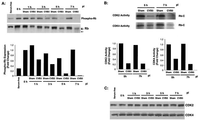
FIG. 2.
CVB3 infection prevents Rb hyperphosphorylation and activation of G1 cyclin kinases. HeLa cells were synchronized by serum starvation, restimulated with serum, and then infected with CVB3 as described in the legend to Fig. 1 or sham infected. (A) Cell lysates were collected and examined by Western blot analysis for hyperphosphorylated Rb (top) and for the levels and relative mobilities of Rb (bottom). Phospho-Rb expression was quantitated by densitometric analysis using NIH ImageJ version 1.27z and normalized to the activity at 0 h p.i., which was arbitrarily set to a value of 1.0. The data represent one of three independent experiments. (B) CDK2 and CDK4 were immunoprecipitated from cell lysates, and kinase activities were determined by an immune complex kinase assay using Rb-C as a substrate. CDK2 and CDK4 activities were quantitated by densitometric analysis and normalized to the sham infection at 1 h p.i. as described above. The data represent one of two independent experiments. (C) Cell lysates were collected, and the expression of CDK2 and CDK4 was examined by Western blotting. The data represent one of three independent experiments.

