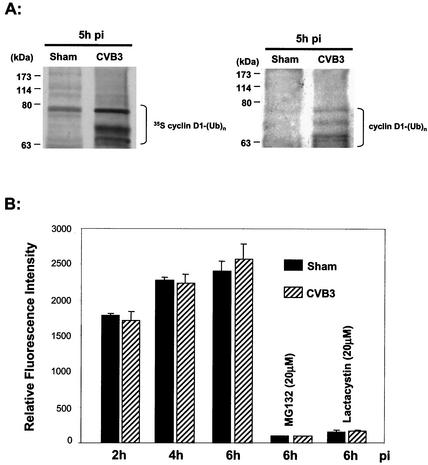
FIG. 6.
CVB3 facilitates ubiquitination of cyclin D1. (A) [35S]methionine-labeled cyclin D1 was analyzed as described in the legend to Fig. 5A. Following separation by SDS-PAGE, the gels were transferred and visualized by autoradiography. On the left is shown the upper portion of the autoradiogram. The same membrane was then examined for ubiquitin expression by Western blotting (right). The masses of protein markers are indicated. (B) 26S proteasome activity following CVB3 infection. At different times after CVB3 or sham infection in the presence or absence of proteasome inhibitors, cell lysates were collected and proteasome activity was measured as described in Materials and Methods using the fluorogenic substrate SLLVY-AMC. The results are means ± SE of three independent experiments.

