FIG. 1.
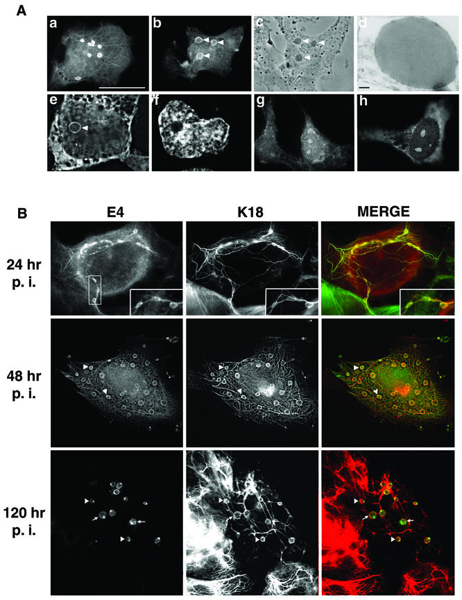
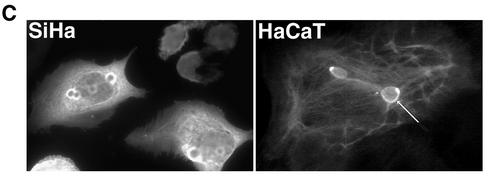
HPV1 E4 accumulates into in vivo-like inclusion bodies in human keratinocytes. (A) Localization of HPV1 E4 in pcDNA-1E4-transfected SVJD keratinocytes. At 48 h after transfection cells were fixed in 4% paraformaldehyde for 5 min prior to acetone permeabilization (10 min) and stained for E4 (MAb 4.37). Immune complexes were visualized with an anti-mouse Alexa Fluor 488 antibody. HPV1 E4 was associated with multiple inclusion bodies that were both phase dense (c, phase-contrast micrograph of cell shown in panel b; inclusions are indicated with arrowheads) and electron dense (d, electron micrograph showing one of the E4 inclusions formed in an Ad-1E4-infected SVJD cell; bar, 100 nm). Inclusions formed in the cytoplasm and nucleus. A deconvolved z section (0.3 μm) of a cell stained for E4 (e) and counterstained with nuclear stain 4′,6′-diamidino-2-phenylindole (Sigma Chemicals) (f) shows a large intranuclear E4 inclusion (arrowhead). In some cells, E4 was also localized to a cytoplasmic filamentous network (a) and small intranuclear foci that resemble nucleoli (g and h). Bar, 20 μm (6.5 μm in panels e and f). (B) Ad-1E4-infected SVJD keratinocytes at a multiplicity of infection of 25 to 50 were fixed at indicated times p.i. in 4% paraformaldehyde for 1 min prior to permeabilization with acetone. Cells were dual stained for E4 (MAb 4.37) and K18 (MAb CK5) and visualized with anti-mouse immunoglobulin G subclass-specific antibodies conjugated to Alexa 488 and Alexa 594 (Molecular Probes Inc.). Deconvolved z-section images are shown in gray and merged (E4 staining is green, K18 staining is red, and colocalization between red and green stains is shown as yellow). Insets in the top panels show enlargement of the marked area and highlight small E4 inclusions interconnected by E4-K18 filaments. In the middle and bottom panels, arrowheads indicate examples of inclusions that show colocalization between E4 and keratin at the periphery of the E4 inclusion. In the bottom panels arrows show E4 inclusions that are surrounded by keratin staining, but the two antigens do not appear to be colocalized. (C) Ad-1E4-infected SiHa and HaCaT keratinocytes (multiplicity of infection of 50) were fixed 48 and 120 h p.i., respectively. An arrow identifies one of the large inclusions formed in the HaCaT cell. HaCaT cells were a gift from N. Fusenig, University of Heidelberg.


