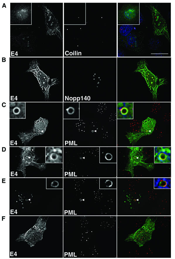
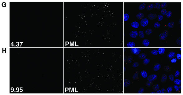
FIG. 4.
Subnuclear topology of HPV1 E4 in SVJD keratinocytes. pcDNA-1E4-transfected SVJD cells were fixed at 48 h in 4% paraformaldehyde for 5 min (A to D and F) or 1 min (E) and permeabilized in acetone. Cells were dual stained for E4 (MAb 4.37) and cellular factors of coilin bodies (A, coilin), nucleoli (B, Nopp140), and ND10 bodies (C to F; PML, rabbit anti-PML antibody). Deconvolved z sections are shown as individual images (gray) and merged (right panels). In merged images, E4 is green; coilin, Nopp140, and PML are red; and where shown 4′,6′-diamidino-2-phenylindole staining of nuclei is blue. Yellow in merged images indicates colocalization between green and red colors. (C to E) Note that in cells containing E4 inclusion bodies PML was reorganized to the periphery of the intranuclear E4 inclusion. The insets in panels C to E are enlargements of the inclusion bodies indicated by arrowheads. PML was partly colocalized with E4. (E) PML-E4 inclusions were also observed in cells that had been fixed differently and stained with a different anti-PML antibody (MAb PG-M3). (F) Note that reorganization of PML did not occur in cells that contain only cytoplasmic E4 inclusions. (G and H) Mock-transfected cells dual stained with 4.37 (G) or 9.95 (H) and PML. Bar, 20 μm (insets, 5.5 μm).


