FIG. 6.
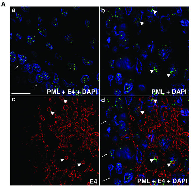
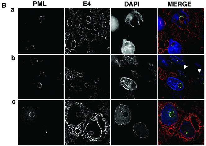
PML is redistributed to intranuclear E4 inclusions in HPV1-induced warts. (A) Tissue sections of wart were dual stained for E4 (MAb 9.95; E4 staining is now shown in red) and PML (rabbit antibody, green), and nuclei were counterstained with 4′,6′-diamidino-2-phenylindole (DAPI) (blue). (a) Merged deconvolved z sections of area of wart negative for E4 staining. Note that PML was localized to multiple intranuclear speckles in basal cells (arrows indicate basal layer) and suprabasal cells. (b to d) PML distribution in area of wart expressing E4. Deconvolved z-section images are shown as PML and DAPI merged (b); E4 alone (c); and E4, PML, and DAPI merged (d). In suprabasal cells, PML was reorganized from multiple speckles to ring structures (b) that are localized to intranuclear E4 inclusions (d; arrows indicate basal cell layer). Arrowheads indicate PML-E4 inclusions. Bar, 20 μm. (B) Reorganization of PML to E4 inclusions was identical to that observed for E4-expressing cultured keratinocytes. Deconvolved z sections are shown as individual images (gray) and merged (E4, red; PML, green; DAPI, blue). PML either nearly completely surrounds the E4 inclusion (a and c) or accumulates predominantly to one side (b). PML shows partial colocalization with E4 (depicted as yellow color in merged images). Note that in panel b the E4-positive cell immediately above the basal layer (indicated by arrowheads) contains a PML-E4 inclusion and that PML distribution is normal in basal cells. Note that the wart section in panel a is stained with MAb 9.95 and that wart sections in panels b and c are stained with anti-E4 MAb 1D11. MAb 1D11 recognizes only full-length E4 (E1^E4) protein. Bar, 10 μm.


