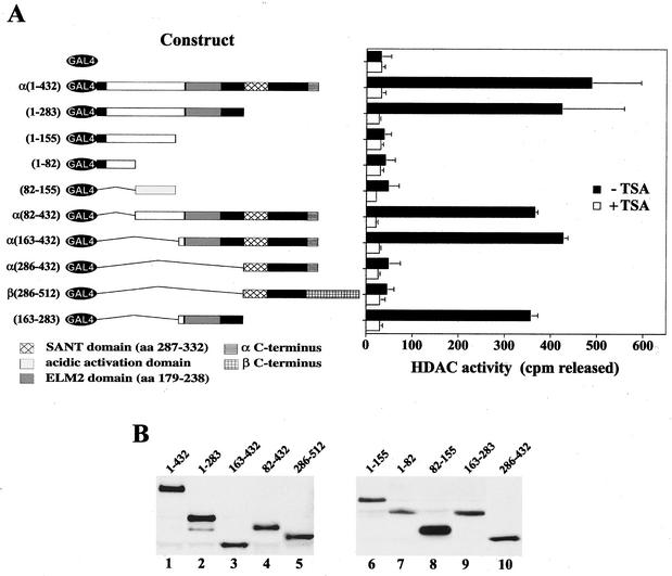
FIG. 4.
hMI-ER1 associates with HDAC activity through a region containing the ELM2 domain. (A) Deletion mutants of hMI-ER1α or β fused to GAL4 were transfected into HeLa cells (1.5 × 10⁵ cells per sample). The schematic on the left illustrates the constructs used and shows a scaled representation of the hMI-ER1 sequence and each of its domains. The individual domains are identified in the legend below the schematic, and the hMI-ER1 amino acid residues encoded by each construct are listed on the left. Cell extracts were prepared 48 h after transfection and subjected to immunoprecipitation with anti-GAL4. Immunoprecipitates were assayed for HDAC activity in the presence or absence of 300 nM TSA as described in Materials and Methods. The histogram shows the average values and standard deviations from three independent experiments. (B) The expression of the GAL4-hMI-ER1 fusion protein in each sample used in panel A was examined by Western blotting using an anti-GAL4 antibody. Indicated above each lane are the hMI-ER1 residues encoded by the construct.

