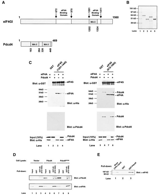
FIG. 7.
Prevention of eIF4A binding to the C-terminal but not to the middle one-third of eIF4G by Pdcd4: Pdcd4 binds to the middle one-third of eIF4G. (A) Structures of eIF4G1 and Pdcd4. The numbers refer to the size (in amino acids) of eIF4G1 and Pdcd4 and to the locations of the eIF4A binding domain and MA-3 domain (2, 32, 40). eIF4A binding domains (open box and arrows) in eIF4G1 are indicated schematically. The MA-3 domains (grey box) in eIF4G1 and Pdcd4 are indicated schematically. (B) Coomassie blue staining of recombinant GST-eIF4G(672-1065), GST-eIF4G(1201-1445), His-eIF4A, and His-Pdcd4. Three micrograms of each recombinant GST-eIF4G(672-1065) (lane 2), GST-eIF4G(1201-1445) (lane 3), His-Pdcd4 (lane 4), and His-eIF4A (lane 5) was resolved by SDS-PAGE and stained with SimpleBlue (Invitrogen). Lane 1, proteinmolecular size markers. (C) In vitro binding assay. Bovine liver GST (lanes 1 and 5), recombinant GST-eIF4G(1201-1445) (lanes 2 to 4), or GST-eIF4G(672-1065) (lanes 6 to 8) was immobilized on glutathione-Sepharose beads and incubated with 5 μg of His-Pdcd4 only (lanes 2 and 6), 5 μg of His-eIF4A only (lanes 3 and 7), or 5 μg of both His-Pdcd4 and His-eIF4A (lanes 1, 4, 5, and 8) on ice for 10 min. After being washed with binding buffer, the bound proteins were resolved by SDS-PAGE and analyzed by immunoblotting with GST antibody (first panel), penta-His antibody (second panel), or Pdcd4 antibody (third panel). Ten percent of input His-Pdcd4 and His-eIF4A proteins were subjected to SDS-PAGE followed by immunoblotting with penta-His antibody (fourth panel). GST-eIF4G(672-1065) and GST-eIF4G(1201-1445) immobilized on glutathione-Sepharose beads were shown as similar amounts. (D) Pulldown of Pdcd4 and Pdcd4D418A with GST-eIF4G(627-1065). JB6 P+ cell lysates isolated following transient transfection with pcDNA3.1+ (lanes 1 to 3), pcDNA-Pdcd4 (lanes 4 to 6), or pcDNA-Pdcd4D418A (lanes 7 to 9) were pulled down with GST (lanes 2, 5, and 8) or GST-eIF4G(627-1065) (lanes 3, 6, and 9). The bound proteins were resolved by SDS-10% PAGE followed by immunoblotting with Pdcd4 or eIF4A antibodies. (E) GST pulldown of endogenous eIF4G with Pdcd4. JB6 P+ cell lysate was pulled down with GST (lane 2) or GST-Pdcd4 (lane 3). The bound proteins were resolved by SDS-10% PAGE followed by immunoblotting with eIF4G antibody. Lane 1 shows one-tenth of the cell lysate.

