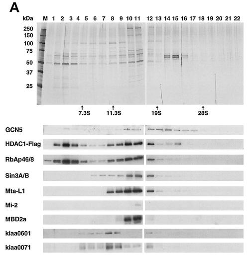
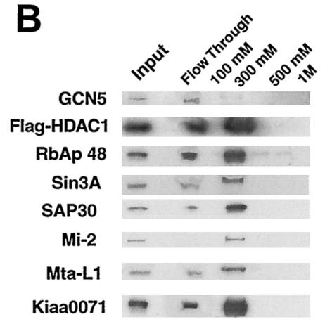
FIG. 4.
Sedimentation analysis of HDAC1-GCN5 complex(es). (A) Using M2 anti-Flag antibody agarose beads, Flag-HDAC1 complexes in HeLa cells were isolated and subjected to 10 to 35% glycerol gradient centrifugation. Each fraction was analyzed on SDS-10% PAGE. Upper two large panels: silver-staining patterns. Markers used were aldolase (7.3S), catalase (11.3S), thyroglobulin (19S), and 28S rRNA, as indicated at the bottom of the two large panels. Lower panels: immunoblot detection of indicated proteins. (B) Flag-HDAC1 complexes were fractionated on a DEAE-Sepharose column and eluted stepwise with buffer containing the indicated NaCl concentrations, and proteins were identified by immunoblotting analysis.


