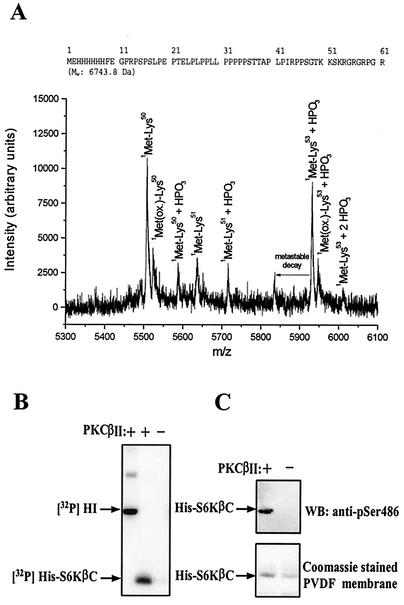
FIG. 3.
Identification of PKC phosphorylation site and characterization of phosphospecific S6Kβ antibody. (A) Mass spectroscopy analysis of PKC phosphorylation site in S6KβII. The amino acid sequence of His-S6KβC is shown on top. (B and C) Analysis of specificity of anti-pS486 antibody. Bacterially expressed His-S6KβC was incubated with [γ-32P]ATP in the presence (+) or absence −) of recombinant PKCβII. Samples were resolved by SDS-PAGE, transferred onto nitrocellulose membranes, and analyzed by autoradiography (B) or immunoblotting with anti-pS486 antibody (C). HI, histone H1; PVDF, polyvinylidene difluoride; WB, Western blot.

