FIG.7.
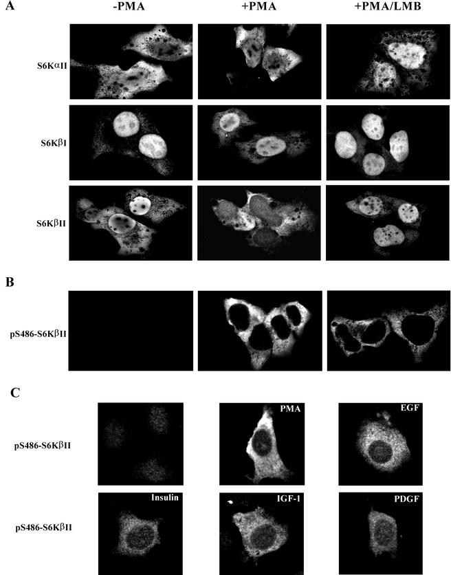
Analysis of subcellular localization of S6Kα and S6Kβ by confocal microscopy. (A) HEK 293 cells were transiently transfected with wild-type EE-S6KαII, EE-S6KβI, or EE-S6KβII, serum starved for 24 h, and stimulated with 1 μM PMA (+PMA) for 30 min or vehicle alone (−PMA). Treatment of cells with LMB (10 ng/ml) was carried out for 16 h before the stimulation with PMA. Cells were fixed, probed with anti-EE antibody and fluorescein isothiocyanate-labeled anti-mouse immunoglobulin G, and analyzed by confocal microscopy. (B) Subcellular localization of pSer486-S6KβII in HEK 293 cells treated with PMA and LMB. HEK 293 cells were transfected with EE-S6KβII and treated in the same way as described above. After fixation and probing with anti-pS486 antibody, confocal microscopy analysis was carried out. (C) Subcellular localization of pSer486-S6KβII in NIH 3T3 cells treated with PMA, EGF, IGF-1, insulin, or PDGF. Transient transfection of NIH 3T3 cells and confocal microscopy were performed as described in Materials and Methods.

