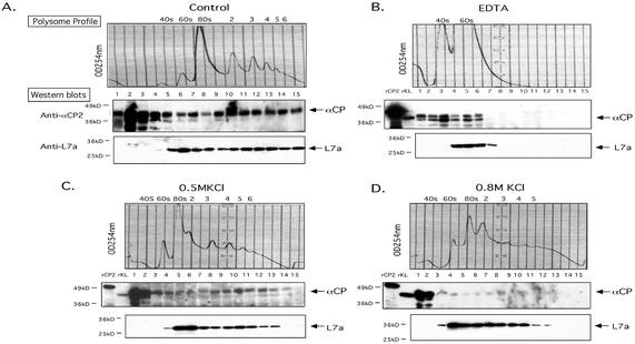
FIG. 4.
Characterization of the αCP-polysome interaction. Polysome aliquots (as in Fig. 3) were treated in the indicated manners and analyzed on 10-to-50% sucrose gradients. (A) No additional treatment. The OD254 is indicated (upper panel; Polysome Profile). Proteins in each fraction were precipitated, separated by SDS-12% PAGE, transferred to membranes, and probed with the indicated antibodies (Western blots). See the legend to Fig. 3 for details. (B) EDTA treatment. The polysome sample was resuspended in 20 mM EDTA (final concentration) prior to sucrose gradient fractionation. This treatment dissociates polysomes into 40S and 60S ribosome subunits. The splitting of the αCP signal seen in this Western blot is occasionally observed. (C) Treatment with 0.5 M KCl. The polysome fraction was brought to 0.5 M KCl (final concentration) prior to sucrose gradient fractionation. This treatment removes proteins loosely associated with the polysomes. (D) Treatment with 0.8 M KCl. The polysome fraction was supplemented with 0.8 M KCl (final concentration) prior to sucrose gradient fractionation. This treatment removes almost all proteins from the polysomes that are not intrinsic ribosomal proteins.

