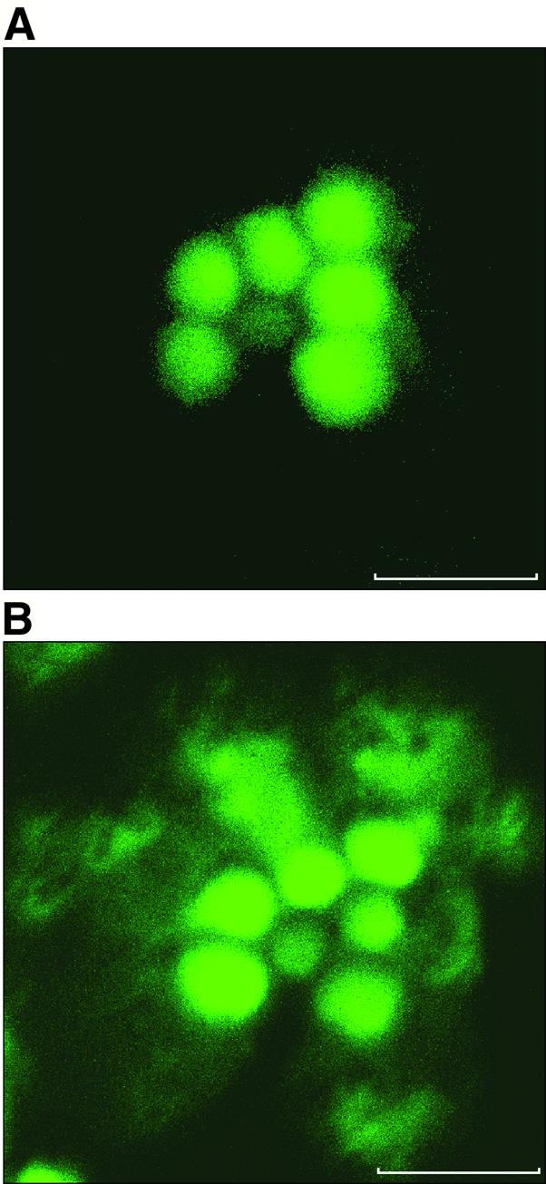
Fig. 3. Light-dependent redistribution of actin in the photoreceptor. Shown are confocal microscopy cross-sections of a single ommatidium in living isolated Drosophila retina. The orientation of the ommatidium is as in Figure 2B. Scale bar = 5 µm. The fluorescence images show GFP–moe marking the actin cytoskeleton (see Materials and methods). (A) Ommatidium of a retina isolated from a dark-adapted fly. (B) Illumination for 60 min results in profound changes in the distribution of the cortical actin to the cytosol outside the rhabdomere, in a pattern resembling that seen in Figure 2E. Similar observations were made in nine different flies.
