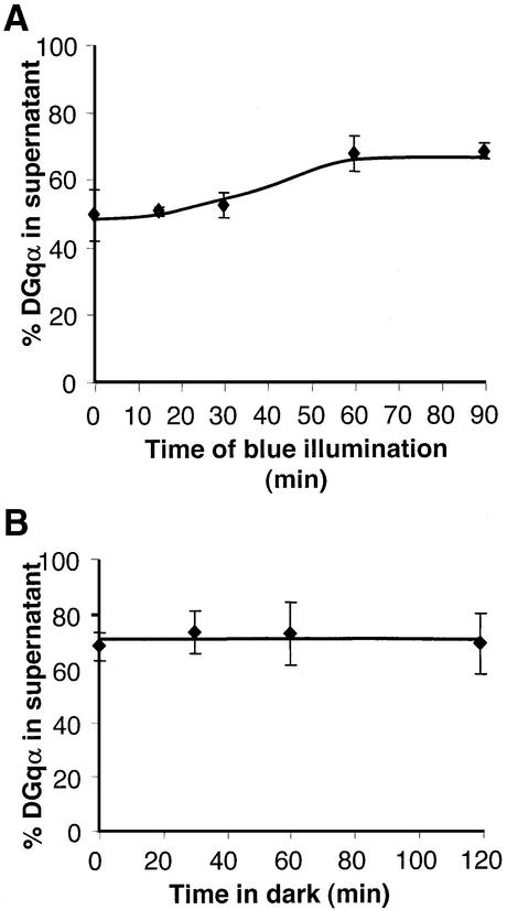
Fig. 7. Severe reduction of Gβ inhibits the translocation of DGqα and abolishes its recovery. (A) The Gβe1 mutant contains low levels of DGqβ. Only 50% of DGqα is localized to the membranes of Gβe1 dark-adapted flies as compared with 80% in wild-type flies (Figure 1). In contrast to the massive initial translocation observed in wild-type flies, in Gβe1 flies there is an obvious lag in the translocation kinetics of DGqα The graph shows the mean amount of DGqα in the supernatant (as a percentage of total) ± SE from four independent experiments. (B) Gβe1 flies were illuminated for 60 min and then incubated in the dark for various times. Even prolonged incubation of Gβe1 flies in the dark (2 h) produced no discernible recovery. The graph shows the mean amount of DGqα in the supernatant (as a percentage of total) ± SE from four independent experiments.

An official website of the United States government
Here's how you know
Official websites use .gov
A
.gov website belongs to an official
government organization in the United States.
Secure .gov websites use HTTPS
A lock (
) or https:// means you've safely
connected to the .gov website. Share sensitive
information only on official, secure websites.
