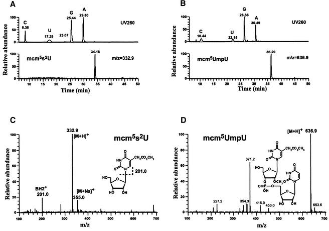
Fig. 2. LC/MS nucleoside analysis of tRNAGlu. (A and B) Chromatograms for cy tRNAGlu and mt tRNAGlu, respectively. Top: UV chromatograms for nucleosides. Bottom: mass chromatograms for modified uridines with mass filters at m/z 332.9 and 636.9 to detect, respectively, mcm5s2U in cy tRNAGlu and a dimer form of mcm5Um with the adjacent uridine (mcm5UmpU) in mt tRNAGlu. (C and D) Mass spectra for (C) mcm5s2U in cy tRNAGlu and for (D) mcm5UmpU in mt tRNAGlu. The chemical structure of each nucleoside is shown within the spectrum.

An official website of the United States government
Here's how you know
Official websites use .gov
A
.gov website belongs to an official
government organization in the United States.
Secure .gov websites use HTTPS
A lock (
) or https:// means you've safely
connected to the .gov website. Share sensitive
information only on official, secure websites.
