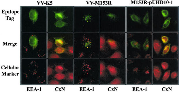
FIG. 5.
Subcellular localization of M153R. HeLa cells were infected with VV-K5 (left panels) or VV-M153R (middle panels) at a multiplicity of infection of 0.1. At 18 h postinfection, cells were stained with antibodies against the cellular marker EEA-1 (early endosomes) or calnexin (CxN) (ER). The right panels show HeLa cells transfected with M153R expressed from expression vector pUHD10-1 (17) and stained as described above. (Bottom row) The binding of primary antibodies was visualized with an Alexa Fluor:568 secondary antibody (red). (Top row) VV-M153R carries a Myc tag and was stained with an FITC-conjugated monoclonal anti-Myc antibody (green). VV-K5 and M153R-pUHD10-1 are Flag tagged. Colocalization is visualized as yellow in the merge panels.

