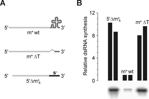
FIG. 8.
The effect of φ6 m+ strand 3′-proximal secondary structure in plus-strand template activity. (A) Diagram of the templates used in the experiment. m+wt, wild-type φ6 m+ segment (T7 transcript from pLM656 cut with XbaI and treated with mung bean nuclease [23]); m+ΔT, m+ segment with deleted cloverleaf structure (T7 transcript from pHY3 cut with BpiI); 5′Δm+s, m+ segment with 3′ terminal part from φ6 s− (T7 transcript from pEM23 cut with BpuAI [16]). (B) The RNAs were incubated with φ6 Pol for 1 h at 30°C as described under Materials and Methods. Reaction products were separated by standard agarose gel electrophoresis and analyzed by phosphorimaging. The autoradiogram shows bands of dsRNA synthesized in each of the three reactions. The graph on the top shows the quantification of the phosphorimager data from two independent experiments.

