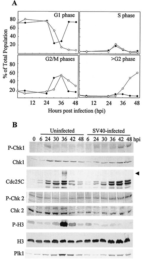
FIG. 1.
Cell cycle progression and expression of MPF regulatory proteins in SV40-infected and uninfected CV-1 cells. Confluent CV-1 cells were infected with SV40 at 100 PFU per cell or trypsinized and replated at 1:3. At 6 hpi or replating, mimosine was added, the cells were incubated for 18 h, and the medium was replaced with fresh medium without mimosine. Samples were harvested at the indicated times. (A) Cells were fixed and stained to determine DNA content per cell by flow cytometry. Uninfected cells, filled circles; SV40-infected cells, open circles. (B) Expression of MPF upstream regulatory molecules and detection of mitotic markers. Whole-cell lysates were resolved by SDS-PAGE and immunoblotted. Each filter was reacted with antibodies specific to each protein and then with alkaline phosphatase-conjugated goat anti-mouse or -rabbit antibody. The arrow indicates hyperphosphorylated (mitotic) Cdc25C.

