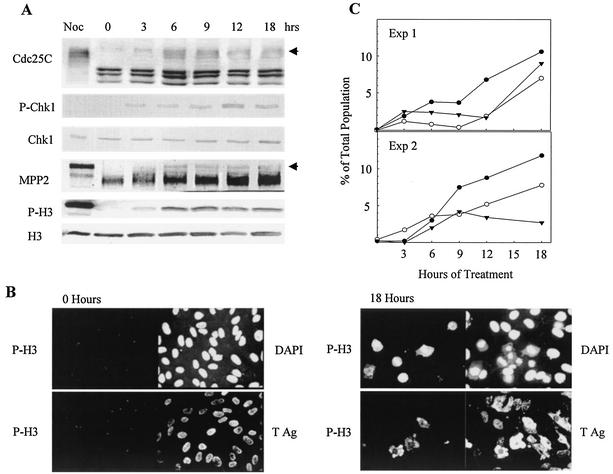
FIG. 3.
Induction of mitotic markers and morphology in SV40-infected cells incubated with UCN-01. Confluent CV-1 cells were infected with SV40 at 100 PFU per cell and treated with mimosine as described in the legend to Fig. 1. At 6 h after release from mimosine, the cells were incubated with 300 nM UCN-01. Samples were harvested at the indicated times postaddition. (A) Induction of mitotic markers. Whole-cell lysates were resolved by SDS-PAGE and immunoblotted. Each filter was reacted with antibodies specific to each protein and then with alkaline phosphatase-conjugated goat anti-mouse or rabbit antibody. Arrows indicate mitotic Cdc25C and mitotic MPP2. The mitotic control was prepared from uninfected CV-1 cells arrested by nocodazole (Noc). (B) Induction of P-H3 in SV40-infected cells. Cells grown on glass coverslips were fixed and stained with anti-P-H3 and anti-large T PAb101 antibodies. Nuclear morphology detail was resolved with DAPI. Cells from all time points were stained, but only samples from the time of addition and the last time point are shown. (C) Nuclear morphology and P-H3 expression. Stained samples of DAPI and P-H3 from panel B were used. DAPI staining was utilized to score the number of cells with normal or abnormal mitotic CC. The number of P-H3-positive cells was also determined. Normal mitotic condensation, filled circles; ACC, open circles; P-H3, filled triangles. Exp, experiment; Ag, antigen.

