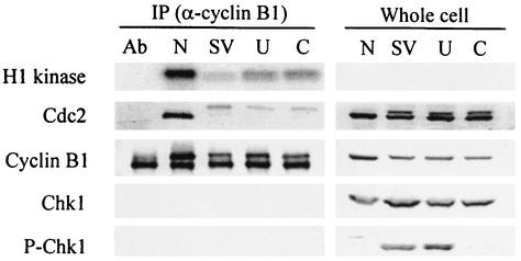
FIG. 6.
Increased MPF protein kinase activity after UCN-01 and caffeine treatments. Confluent CV-1 cells were infected with SV40 at 100 PFU per cell and treated with mimosine as described in the legend to Fig. 1. Caffeine (6 mM) was added to one set of cultures at 3 h after release from mimosine (lane C). UCN-01 (300 nM) was added to one set of cultures at 6 h after release from mimosine (lane U). A parallel infected culture released from mimosine was not treated with any mitotic inducers (SV). Samples were harvested at 48 hpi. A mitotic control was prepared from uninfected CV-1 cells arrested by nocodazole (lane N). Lysates were precipitated with mouse anti-cyclin B1 antibody (Ab). Precipitated samples (2 × 10⁶ cells) were used for a kinase assay with histone H1 as the substrate. Phosphorylated H1 was resolved by SDS-PAGE and visualized by phosphorimaging. The remaining precipitated samples and whole-cell lysates were resolved by SDS-PAGE and immunoblotted. Each filter was reacted with antibodies specific to each protein and then with alkaline phosphatase-conjugated goat anti-mouse or -rabbit antibody. An anti-cyclin B1 immunoprecipitation without cell extract is shown in lane Ab.

