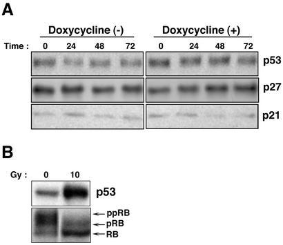
FIG. 6.
Expression levels of p53 and CDK inhibitors, p21WAF-1/CIP-1 and p27KIP-1, after induction of the lytic program in Tet-BZLF1/B95-8 cells. Tet-BZLF1/B95-8 cells were cultured in the presence (+) or absence (−) of 1 μg of doxycycline/ml and harvested at the indicated times (in hours). Clarified cell lysates were prepared, separated by SDS-10% PAGE (p53 and CDK inhibitors) or by SDS-7.5% PAGE (pRB), and subjected to Western blot analysis with each specific antibody. Corresponding proteins were detected with enhanced chemiluminescence reagents. The images were processed by LumiVisionPRO (Aisin/Taitec Inc.) with a cooled CCD camera and assembled in an Apple G4 computer using Adobe Photoshop version 5.0. The signal intensity was quantified with a LumiVision image analyzer. (A) Expression levels of p53, p21WAF-1/CIP-1, and p27KIP-1 in Tet-BZLF1/B95-8 cells after doxycycline annexation. Tet-BZLF1/B95-8 cells were untreated or treated with doxycycline (1 μg/ml) and harvested at the indicated times. The expression level of p16INK4A was low and was not affected by the addition of doxycycline (data not shown). (B) Levels of p53 and phosphorylation status of Rb protein by gamma irradiation. Tet-BZLF1/B95-8 cells were irradiated with gamma radiation at a dose of 10 Gy and harvested 6 h postirradiation. The slower-migrating bands are hyperphosphorylated forms of the Rb protein (pRB and ppRB). The faster-migrating band is the hypophosphorylated form of the Rb protein (RB).

