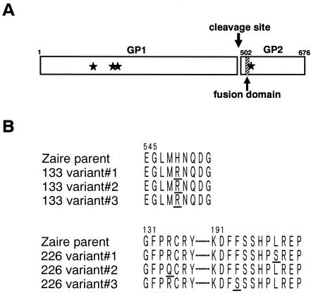
FIG. 5.
(A) Schematic diagram of Ebola virus GP. Ebola virus GP is proteolytically cleaved into GP1 and GP2 subunits (20). The fusion domain, a highly conserved hydrophobic region (amino acids 524 to 539), is located 24 amino acids downstream of the N terminus of the GP2 subunit. Stars represent the positions of amino acid substitutions. (B) Amino acid substitutions found in escape mutants selected by MAbs 133/3.16 (133 variants 1 to 3) and 226/8.1 (226 variants 1 to 3). Substituted amino acids are underlined.

