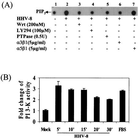
FIG. 2.
HHV-8 activates PI 3-kinase in the target cells via its interactions with α3β1 integrin. (A) Induction of PI 3-kinase by HHV-8. Serum-starved HFF cells were mock infected (lane 1) or infected with HHV-8 (lane 2) for 5 min. Cells were also preincubated with the PI 3-kinase inhibitors wortmannin or LY294002 for 1 h at 37°C and then infected with virus in the presence of inhibitors for 5 min (lanes 3 and 4). The phosphotyrosine residue in PI 3-kinase was dephosphorylated by incubating the immunoprecipitates with 0.5 U of PTPase 1B for 2 h at 30°C before an assay for PI 3-kinase activity (lane 5) was carried out. Virus was also preincubated with 5 μg of soluble α3β1 or α5β1/ml for 1 h at 37°C before infection of the cells (lanes 6 and 7). Equal protein concentrations of cell lysates were immunoprecipitated with PY20 antibodies, assayed for PI 3-kinase activity by using sonicated PIP2 as the substrate, and resolved with radiolabeled PIP3 by ascending thin-layer chromatography, and PIP3 spots were detected by autoradiography. (B) Kinetics and quantitation of PI 3-kinase activity induced by HHV-8. Serum-starved HFF cells were mock infected for 15 min or infected with HHV-8 for the indicated time points or treated with FBS for 15 min and assayed for PI 3-kinase activity as described in Fig. 2A. Radioactive PIP3 spots were quantitated and expressed as fold change of PI 3-kinase activity over mock-infected cells.

