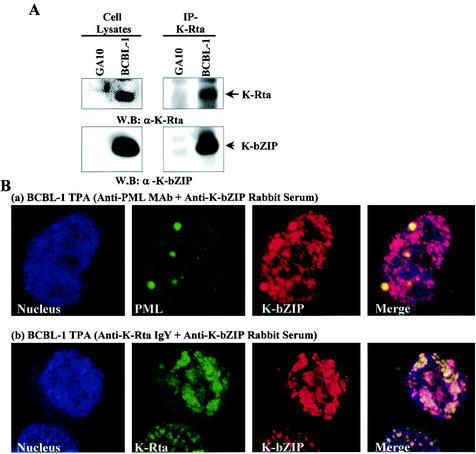
FIG. 1.
Coimmunoprecipitation assay. (A) Coimmunoprecipitation assay with the KSHV-positive BCBL-1 cell line. The GA-10 cell line was used as a KSHV-negative B-cell control. BCBL-1 cell lines induced with TPA (48 h) were harvested, and the same amounts of lysates (1.0 mg) were precipitated with anti-K-Rta chicken IgY and then immunoblotted with anti-K-bZIP rabbit antibody. IP, immunoprecipitation; W.B, Western blotting; α, anti. (B) Colocalization of K-bZIP with PML (a) and K-Rta (b) in BCBL-1 cells. Confocal analyses were performed by using anti-K-bZIP rabbit serum and anti-PML mouse monoclonal antibody (MAb) or anti-K-Rta chicken IgY. K-bZIP (red), PML (green), and K-Rta (green) were detected with rhodamine-conjugated anti-rabbit IgG and FITC-conjugated anti-mouse IgG or anti-chicken IgY. The nucleus was counterstained with TO-PRO-3 (blue). These panels are representative of 10 different fields.

