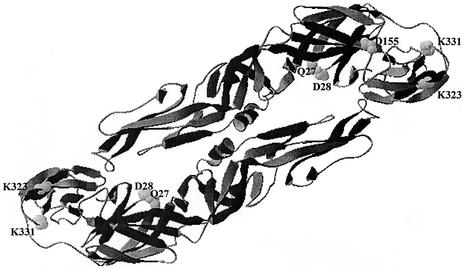
FIG. 6.
Three-dimensional model of the YF virus E protein based on the crystallographic structure of TBE virus E protein (26). The five amino acid positions that differ between the Asibi/hamster p0 and Asibi/hamster p7—Q27, D28, D155, K323, and K331—are highlighted and labeled. The Swiss PDB program was used to model the YF virus E protein onto the crystallographic structure of the TBE virus E protein. The orientation of the monomers to form a dimer was approximated.

