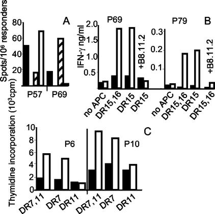
FIG. 3.
HLA allele-specific presentation of peptides. (A) IFN-γ Elispot assay with PBMC of donor VB-7 (DRB1*1101,*1501) depleted of CD8- and HLA class II-positive cells. As antigen-presenting cells, we used PBMC from homozygous donors (VB-10, DRB1*1101, and VB-12, DRB1*1501) that were depleted of CD3-positive cells. Black bars, VB-7 PBMC not depleted; white bars, DRB1*1101 antigen-presenting cells; hatched bars, DRB1*1501 antigen-presenting cells. From the number of spots depicted, the background in the absence of peptide has been subtracted. (B) IFN-γ production of short-term T-cell lines. The responder populations were incubated for 3 days with heterozygous (CH-1, DRB1*1501, -*16) antigen-presenting cells or homozygous (VB-12, DRB1*1501) antigen-presenting cells. Both antigen-presenting cell populations were CD3 depleted. Black bars, no peptide; white bars, 20 μM peptide 69 (left side) or 20 μM peptide 79 (right side). B8.11.2, anti-HLA-DR antibody. (C) Proliferation assay with short-term T-cell lines of donor VB-2 (DRB1*0701, *1101). Antigen-presenting cells were from VB-8 (DRB1*0701) or VB-10 (DRB1*1101). Black bars, no peptide; white bars, 20 μM peptide 6 (left side) or 20 μM peptide 10 (right side).

