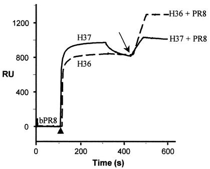
FIG. 4.
Valency of binding of H37 IgG applied at high concentration: a proportion of this IgG that bound bivalently at low concentration now binds monovalently. The baseline represents bound bPR8 (Fig. 2). The first part of each curve shows the injection of 160 μg of H37 and H36 IgGs per ml (arrowhead) and their binding to immobilized bPR8, under the conditions described in the Fig. 2 legend. After the plateau, there is some dissociation of H37 IgG. After the injection of detector nonbiotinylated PR8 (arrow), there is an increase in RU due to its capture by antibody. H37, solid line; H36, dashed line.

