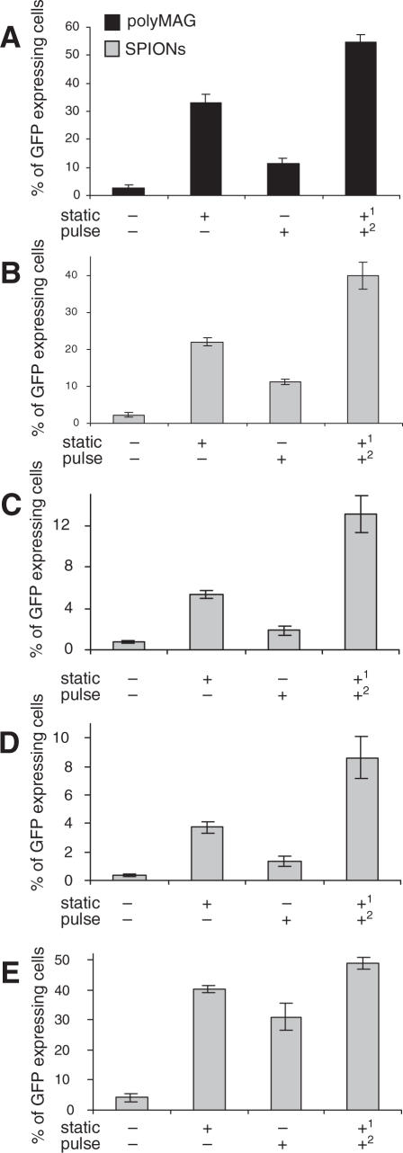
Figure 3.
Effect of pulsating magnetic field on expression of GFP. The 293T cells were transfected with either DNA/polyMAG (A) or DNA/SPIONs (B), and placed either on a static magnetic plate for 5 min, or a pulsating magnetic field was applied for 5 min, or the pulsating field was applied after the static field. (C) HeLa cells, (D) Cos7 cells and (E) synoviocytes were transfected with DNA/SPIONs as stated above. Control cells were not exposed to any magnetic field. Numbers 1 and 2 represent the application sequence of the magnetic fields. Results are shown as means and SD values from at least three independent experiments.

