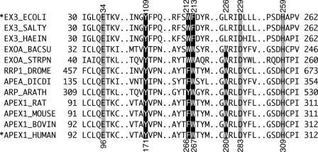
Figure 1.
Sequence alignment of active sites of AP endonucleases. The amino acid sequences of AP endonuclease were aligned using EX3_ECOLI (E.coli ExoIII), EX3_SALTY (Salmonella typhimurium ExoIII), EX3_HAEIN (Haemophilus influenzae ExoIII), EXOA_BACSU (B.subtilis ExoA), EXOA_STRPN (Streptococcus pneumoniae ExoA), RRP1_DROME (Drosophila melanogaster Rrp1), APEA_DICDI (Dictyostelium discoideum APEA), ARP_ARATH (Arabidopsis thaliana ARP), APEX1_RAT (Rattus norvegicus APE1), APEX1_MOUSE (Mus musculus APE1), APEX1_BOVIN (Bos taurus APE1) and APEX1_HUMAN (H.sapiens APE1). The reversed and the shaded characters indicate conserved aromatic amino acid residues and active amino acid residues, respectively. The amino acid residue numbers are given at the left and right of each line. The corresponding numbers of the amino acid residues in EX3_ECOLI and APEX1_HUMAN are shown above and below the sequences, respectively. Asterisks indicate the AP endonucleases used in this study.

