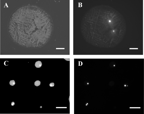
Figure 2. Optical micrographs of droplet relics containing CAD cells.
The optical micrographs shown here were obtained with phase-contrast and fluorescence optics. Cell suspensions containing Rhodamine-stained CAD cells in fluorescein-labelled medium were jetted at 10 kV on to glass slides (A and B) or on to nylon membrane (C and D). (A) shows a dried droplet relic seen under phase-contrast, and (B) shows the same relic using Rhodamine filters, demonstrating that two cells were present in the droplet. The scale bars in (A) and (B) represent 100 μm. (C) and (D) show an example of droplet relics after the cell suspension was jetted on to nylon membrane. (C) shows a group of spots observed using fluorescein filters and (D) shows the same spots using Rhodamine filters. The scale bars for (C) and (D) represent 500 μm.

