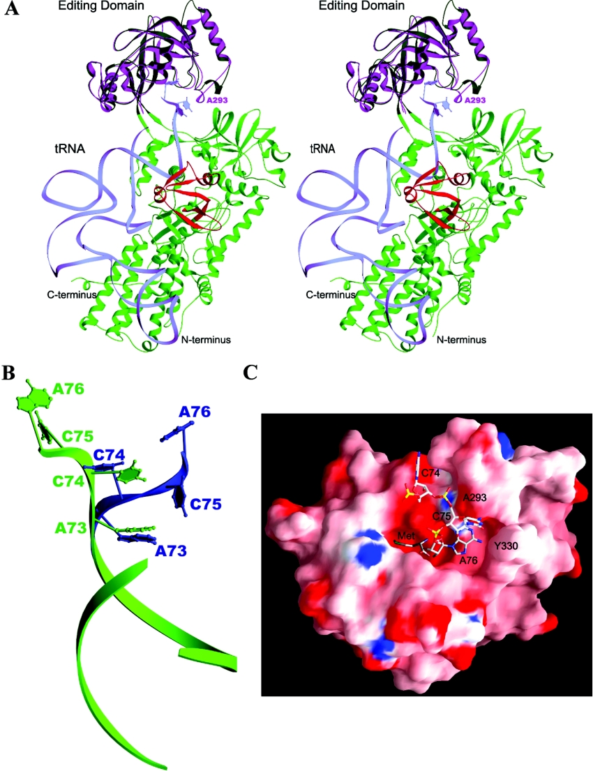
Figure 4. Docking model of the ecLeuRS-ED–tRNA complex.
(A) The docking model of ttLeuRS in complex with tRNA. The subdomains of ttLeuRS are coloured as the RF domain and the small helical domain in green, ED in dark green, and LeuRS specific domain in red. The structure of ecLeuRS-ED (in purple) is superimposed onto the editing domain of ttLeuRS. tRNA is shown as light blue ribbon and the 3′ terminal CCA are shown with nucleotide bases. (B) Comparison of the 3′ terminal ACCA of the tRNA between the ValRS–tRNA complex (in blue) and the docking model of the ecLeuRS-ED–tRNA complex (in green). (C) Molecular surface of the Met-bound ecLeuRS-ED structure viewing from the entrance to the editing active site. Both the bound Met substrate and the docked 3′ terminal CCA of the tRNA are shown as ball-and-stick models.

