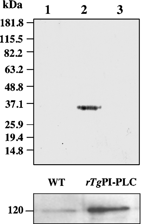
Figure 7. Biotinylation of cell-surface proteins in tachyzoites.
Upper panel, tachyzoites of the 2F1 strain were incubated with 2 mM sulpho-NHS-biotin for 30 min. After lysis of the cells with RIPA buffer, the lysates were immunoprecipitated by the affinity-purified anti-TgPI-PLC polyclonal antibody, and the immunoprecipitates were subjected to Western blot analysis. Detection of biotinylation was carried out using streptavidin–peroxidase conjugate and ECL®. No band was detected in the immunoprecipitates with anti-PI-PLC (lane 1). Lane 2 shows a positive control with anti-SAG1 antibody instead of anti-TgPI-PLC antibody for immunoprecipitation. Lane 3 shows a negative control with anti-β-galactosidase antibody for immunoprecipitation. Migration of molecular-mass standards (in kDa) is shown to the left of the gels. Lower panel, the positive control experiment shows that the anti-TgPI-PLC antibody can immunoprecipitate both the native PLC from T. gondii (left-hand lane) and the recombinant TgPI-PLC (right-hand lane), as probed with the guinea-pig anti-TgPI-PLC antibody. The position of a 120 kDa protein is indicated.

