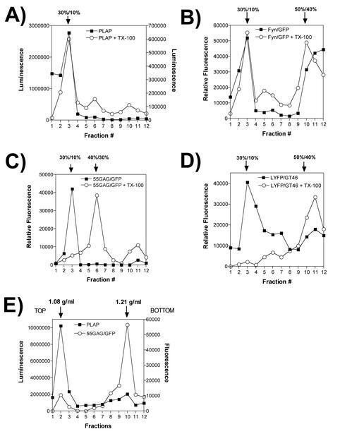
FIG. 3.
Independent segregation of Gag DRCs and lipid raft markers. Proteins were expressed and cells were processed in an identical manner to the proteins and cells shown in Fig. 1 to 3, and postnuclear supernatants were subjected to equilibrium flotation on 50%/40%/30%/10% iodixanol gradients. Flotation results obtained from samples extracted with TX-100 (open circles) and results obtained without detergent treatment (closed squares) are shown. All processing was performed on ice or at 4°C. (A) PLAP activity was assessed using a chemiluminescence assay for alkaline phosphatase activity. (B) Fyn-GFP quantitation by fluorometry. (C) 55GAG/GFP fluorescence profile before and after TX-100 extraction. (D) LYFPGT46, a nonraft transmembrane marker protein, was quantitated by fluorometry in the absence of detergent or following TX-100 treatment. (E) Separation of raft markers and Gag-GFP was performed on 20 to 75% sucrose gradients. The density of peak fractions was measured by refractometry and is indicated above the graph.

