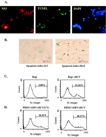
FIG. 8.
HCV infection causes apoptosis in HCV-infected B cells. (A) SB cells were costained with anti-HCV NS3 antibody, fluorescence-labeled deoxynucleoside triphosphate for TUNEL assay, and DAPI for visualization of all cells. Most of the TUNEL-positive cells were also positive for NS3. (B) Detection of apoptotic cells by TUNEL assay in uninfected Raji cells (left) and HCV-infected Raji cells (right). Apoptotic cells were stained brown. The percentage of apoptotic cells was determined as the average of four fields under the microscope. This experiment was repeated five times. The range of percentages of apoptosis was 1 to 5% for Raji cells and 21 to 29% for HCV-infected Raji cells. (C) Analysis of apoptotic cells by fluorescence-activated cell sorter in HCV-infected (Raji + HCV) and uninfected Raji cells by annexin V-binding assay. (D) Analysis of apoptotic-cell populations in PBMCs coinfected with EBV and SB culture supernatant (HCV) or UV-irradiated SB supernatant [HCV (UV)]. The assays were done on the cells 2 months postinfection.

