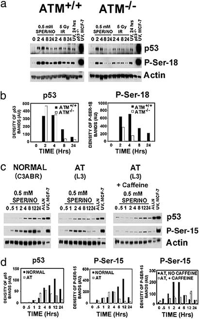
Figure 2.
(a) ATM partially mediates NO-induced P-Ser-18 in MEFs. ATM+/+ or ATM−/− MEFs were exposed to 0.5 mM SPER/NO, 5 Gy γ-irradiation, or 25 J/m2 UV for indicated time points (hr). Cells were lysed, and Western blot assays were performed. (b) Bar graphs representing quantitative densitometry of Western blot bands shown in a. (c) ATM and ATR mediate NO-induced P-Ser-15 in human cells. Human lymphoblastoid cells from a healthy individual (C3ABR) or an individual with ataxia telangiectasia (AT) were exposed to 0.5 mM SPER/NO ± caffeine (1 mg/ml) for indicated time points (hr). The calpain inhibitor, ALLN (20 μM), which inhibits the proteasome, was used as a negative control for posttranslational modifications; MCF-7 cells exposed to 25 J/m2 UV were used as a positive antibody control. Cells were lysed, and Western blot assays were performed. (d) Bar graphs representing quantitative densitometry of Western blot bands shown in c.

