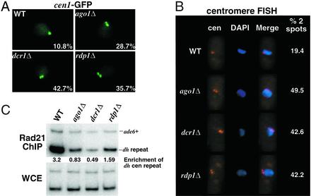
Figure 2.
Sister chromatid cohesion at centromeres is disrupted in the RNAi mutant strains. (A) Localization of cen1 in live cells as visualized by accumulation of LacI-GFP at the LacO array inserted at the lys1 locus linked to cen1. GFP spots were counted on a computer screen after capturing serial images of fields of cells at 0.4-μm intervals along the z axis. Spots were deemed distinct when their midpoints were separated by a distance greater than or equal to their respective radii. The percentage with which each genotype displayed two GFP spots is noted in the lower right corner of each image. More than 100 cells were counted for each strain. (B) FISH analysis of wild-type and mutant cells using a 15-kb probe that hybridizes to the outer repeats of all three centromeres. The number of spots were counted by microscopic inspection of >100 cells for each strain. (C Upper) ChIP analysis of Rad21-HA in wild-type and mutant strains using the 12CA5 antibody. Relative fold enrichments of dh centromeric repeats are indicated beneath each lane. (Lower) DNA prepared from whole cell extracts (WCE).

