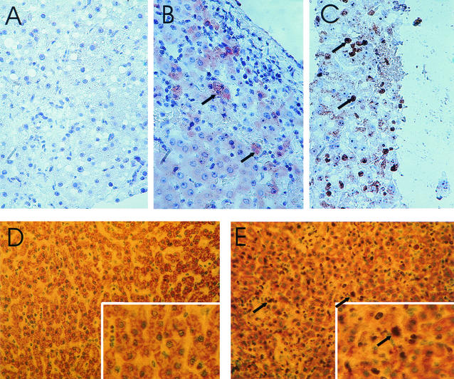
FIG. 1.
Liver sections from rhesus macaques infected with LCMV-WE. (A and B) Immunoperoxidase staining of necropsy samples from healthy monkey Rh-ig6a (A) and lethally infected monkey Rh-iv6 (B). LCMV viral antigen (red) in hepatocytes of Rh-iv6 is indicated with arrows. Magnification, ×200. (C) Biopsy sample, taken 4 weeks after infection, from transiently ill monkey Rh-ig7b during the onset of illness. Brown staining of hepatocyte nuclei (arrows) indicates positivity for proliferation antigen Ki-67. Liver biopsies taken before infection and stained with Ki-67 looked like the picture seen in panel A. Magnification, ×300. (D and E) Ki-67 staining of necropsy liver tissue in healthy monkey Rh-ig6a (D) and in fatally infected monkey Rh-iv6 (E). Note brown staining of Ki-67-positive nuclei (arrows) in panel E. Magnifications, ×100 (main panels) and ×300 (insets).

