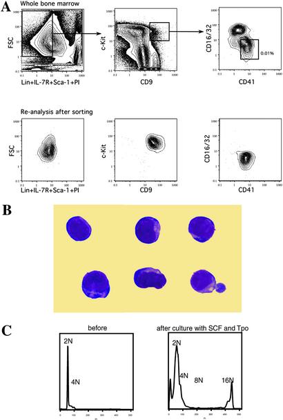
Figure 1.
Identification of MKPs in mouse bone marrow. (A) Flow cytometric analysis of bone marrow after depleting the lineage-positive cells with magnetic beads. (Upper) The sorting gates for MKPs. The frequency of MKPs is shown as relative to total nucleated bone marrow cells before the negative depletion. Reanalysis after the first round of sorting found MKPs to be cleanly isolatable population (Lower). (B) MKPs were myeloblast-like cells with no characteristic features of megakaryocytes (Giemsa staining, original magnification ×1,000). (C) DNA content analysis showed MKPs to be diploid cells. After a 5-day incubation with SCF and Tpo, polyploid (16N) megakaryocytes were found in the culture.

