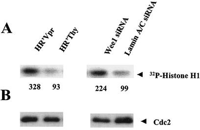
FIG.3.
Cdc2 kinase activity is increased in HR′Vpr and Wee-1 siRNA-transfected HeLa cells. Cells were infected as described in the legend for Fig. 1 and transfected in parallel with those shown in Fig. 2. Cells were collected at 72 h post-virus infection or 24 h post-siRNA transfection. (A) Kinase assay of Cdc2. The assay was performed as described in Materials and Methods. The results were visualized and quantified using a PhosphorImager (Molecular Dynamics), and the numbers represent the relative density of the 32P-labeled histone H1. (B) Cdc2 protein level the kinase assay shown in panel A. Five micrograms of cell lysates was fractionated by SDS-12% PAGE and immunoblotted with Cdc2 antibody as described in the legend for Fig. 1. Relative Cdc2 activities were determined after normalization with the Cdc2 protein amount.

