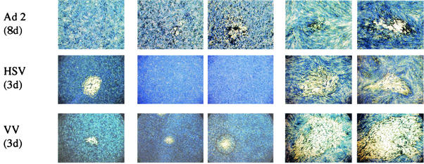
FIG. 3.
Photographs of plaques of a variety of RNA and DNA viruses on Vero, HEp2, HEp2/SV5-V, MRC5, and MRC5/SV5-V cells grown in six-well petri dishes. The cells were fixed at various days p.i. (indicated in parentheses) and stained with Coomassie brilliant blue. Photographs were taken on an inverted microscope at 4× magnification. The viruses used in the plaque assays were measles virus (MeV, a wt isolate), canine distemper virus (CDV), SV5 (strain W3), hPIV2 (a wt and a recent clinical isolate, 5234), mumps virus (a wt and the Enders strain [att]), hPIV3 (PIV3), Theiler's virus, adenovirus type 2, HSV (strain STH2), and vaccinia virus (VV).


