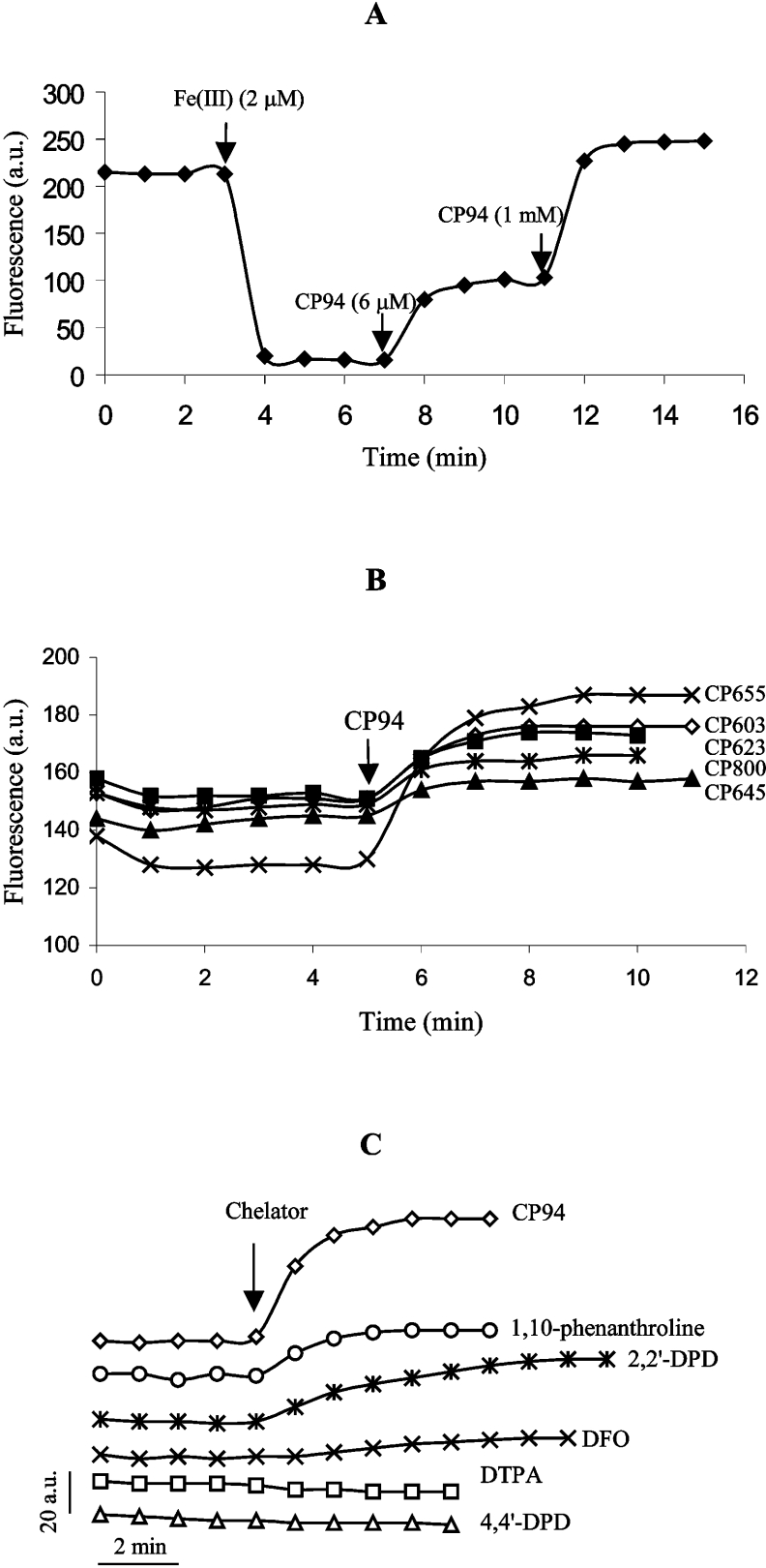
Figure 3. Evaluation of fluorescence dequenching of pyridinone fluorescent probes.
(A) Fluorescence (λex=416 nm, λem=474 nm) of CP655 (6 μM) was quenched by the addition of 2 μM iron(III) and then recovered after addition of 6 μM and 1 mM CP94. (B) Effect of CP94 (1 mM) on the intracellular fluorescence of hepatocytes that had been loaded with various probes (10 μM each). (C) Effect of various chelators [CP94, 1,10-phenanthroline, 2,2′-dipyridyl (2,2′-DPD), DFO and DTPA; 1 mM each] and the non-chelating analogue of 2,2′-dipyridyl, 4,4′-dipyridyl (4,4′-DPD) (1 mM), on hepatocellular CP655 fluorescence.

