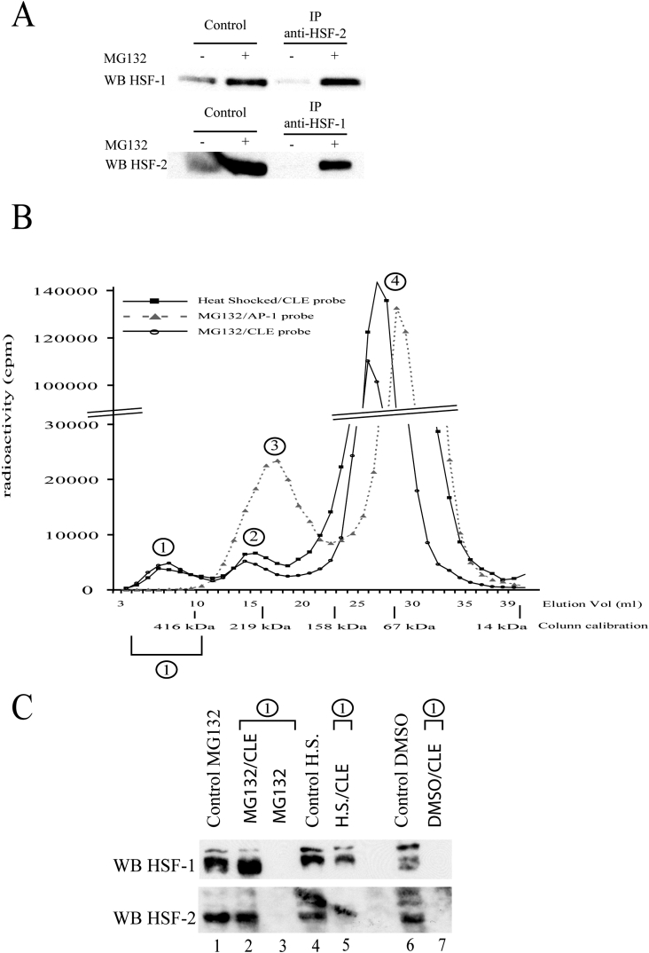
Figure 5. HSF1 and HSF2 form a heterocomplex under MG132 treatment.
(A) HSF1 and HSF2 co-immunoprecipitations. Cell extracts from untreated (−) or MG132-treated (+) cells (5 μM, 16 h) were used to detect HSF1 or HSF2 protein by Western blotting, before (control) or after immunoprecipitation (IP) with specific anti-HSF1 or anti-HSF2 antibodies. The Figure presented is representative of three independent experiments. (B) Estimation of the relative size of HSF–CLE complexes. 32P-labelled CLE or AP-1 probes were incubated with nuclear extract from U-251 MG cells treated as indicated. The sizes of the DNA–protein complexes were estimated by gel-filtration chromatography on a Superdex 200 column, which was calibrated using different globular proteins. Presence of DNA probe was determined by quantification of radioactivity (c.p.m.) in each fraction. Peak 1 corresponds to the specific HSF–CLE complex, whereas peaks 2, 3 and 4 correspond to non-specific complex, AP-1-binding complex and free probes respectively. Identities of each peak were determined by carrying out gel filtration with labelled DNA probe alone (peak 4), or with probe previously incubated with nuclear extract in the presence of high excess of non-specific DNA (peak 2 affected), or with high excess of specific CLE unlabelled competitor (peak 1 disappeared). Breaks in the y-axis scale are for better readability of the Figure. (C) Western-blot analysis of peak 1 contents. Western-blot analyses were carried out on nuclear extracts, before (control, lanes 1, 4 and 6) or after gel-filtration chromatography (lanes 2, 3, 5 and 7). Extracts from untreated cells (DMSO; lanes 6 and 7), MG132-treated cells (lanes 1–3) or from heat-shock-treated cells (H.S., lanes 4 and 5) were used. Gel filtration was performed as described in the Materials and methods section, in the presence (lanes 2, 5 and 7) or absence (lane 3) of CLE probe. Elution fractions corresponding to peak 1 were then pooled, precipitated and assayed for HSF1 and HSF2 presence using specific anti-HSF1 and anti-HSF2 antibodies (upper and lower panels respectively). An unspecific stain is visible in the lower panel, overlapping lanes 4 and 5.

