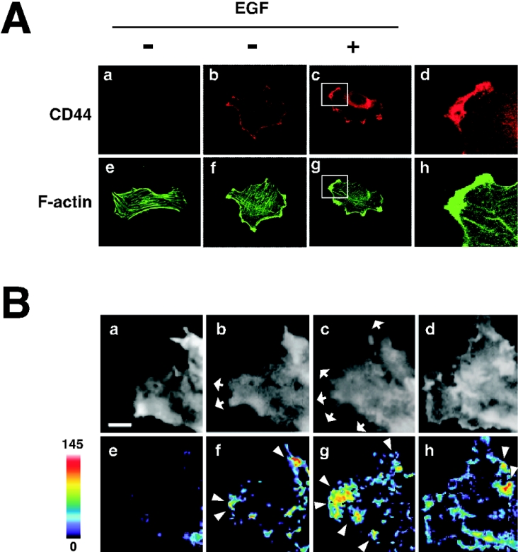
Figure 2. EGF induces redistribution of CD44 to the lamellipodial membrane protrusion area and peripheral activation of Rac1.
(A) Immunocytochemical staining of CD44. U251MG cells incubated with (panels c and g) or without (panels a, b, e and f) 10 ng/ml EGF were stained with anti-CD44cyto pAb (panels b and c) or a control antibody (panel a). The cells were also stained with Alexa Fluor® 488-conjugated phalloidin (panels e, f and g). Panels d and h are enlargements of the regions indicated in panels c and g respectively. (B) In situ detection of Rac1 activation. U251MG cells were transfected with YFP–Rac1 followed by microinjection with BODIPY® TR–GTP. The cells were treated with 10 ng/ml EGF, and the images after 0 (panels a and e; before EGF treatment), 5 (panels b and f), 10 (panels c and g) and 30 min (panels d and h) were acquired through the YFP (panels a–d) and the FRET (panels e–h) channels under confocal microscopy. FRET signal intensity from 0 to 145 is displayed using pseudocolour as indicated by the scale bar. Arrowheads indicate the areas where intense FRET signal was detected, and arrows show the areas of outgrowth. Scale bar, 2 μm.

