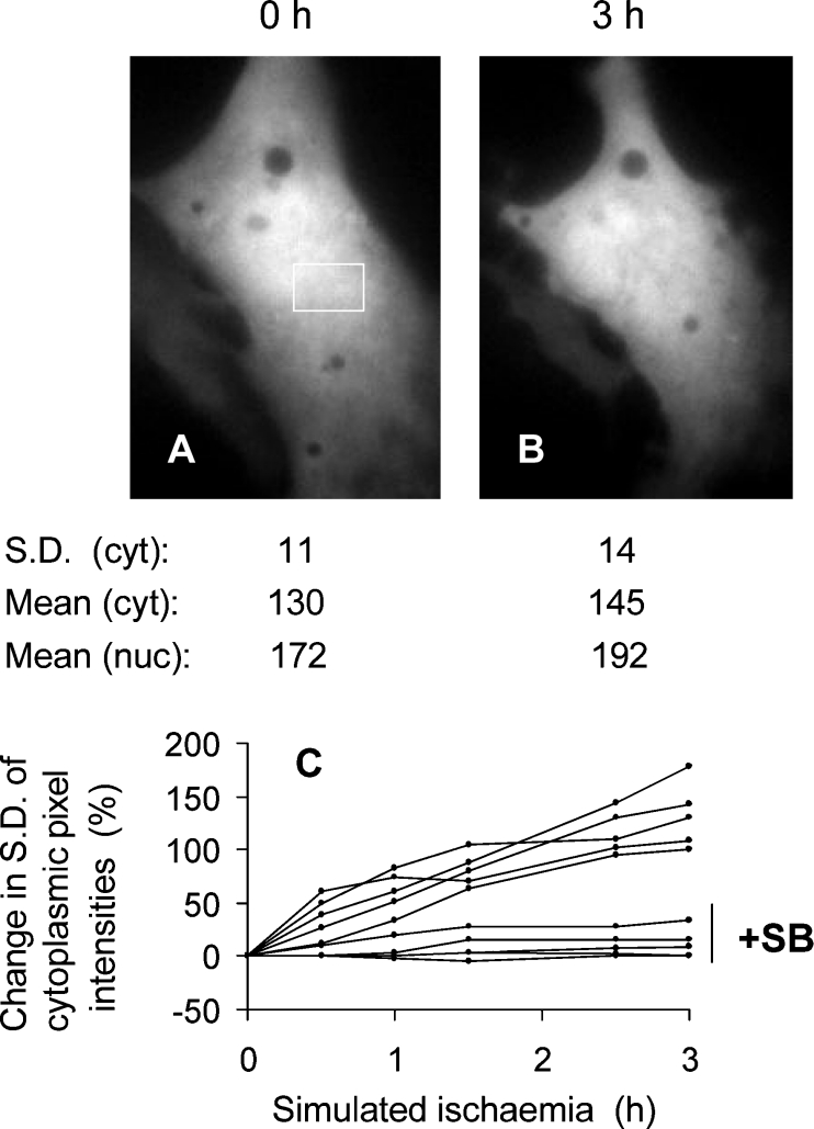
Figure 4. The lack of GFP–Bax sequestration during simulated ischaemia in the presence of SB203580.
A GFP–Bax-transfected cell was preincubated in standard medium containing 10 μM SB203580 and imaged (A). The medium was then replaced with glucose-free medium containing CN and SB203580, and the cell was imaged 3 h later (B). The S.D. of cytoplasmic (cyt) pixel intensities refers to the area boxed in (A). The mean nuclear (nuc) and cytoplasmic fluorescence values were obtained after defining the boundaries from brightfield images. (C) Time courses of GFP–Bax sequestration according to the changes in S.D. of cytoplasmic pixel intensities. Each line was from a single cardiomyocyte in the presence or absence of 10 μM SB203580 (SB), as indicated.

