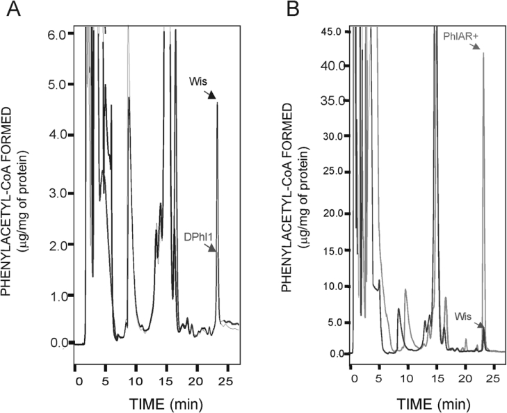
Figure 5. PAA-CoA formed in vitro by the PAA-CoA ligase.
(A) PAA-CoA formed in vitro by the PAA-CoA ligase of the parental strain Wis 54-1255 and the disrupted DPhl1. (B) PAA-CoA formed in vitro by the PAA-CoA ligase of the parental strain Wis 54-1255 and the PhlAR+ transformant containing multiple copies of the phl gene. Pure PAA-CoA was used as standard (elution time 23.5 min). A stock solution of 100 μg/ml PAA-CoA was used to determine the standard curve. The reactions were performed with 500 μg of protein (crude extract) per reaction (0.3 ml volume) in all cases. Note the different scales between panels (A) and (B).

