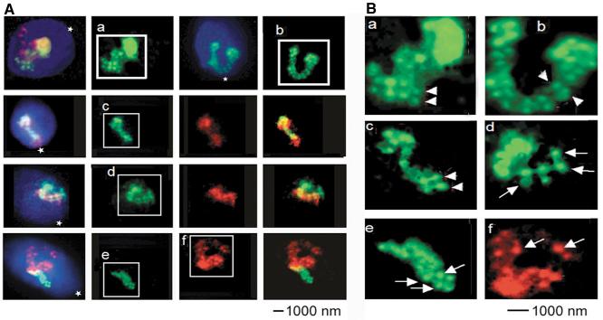
Fig. 4.
Internal organization of chromosome arm fiber. (A) Representative images of CHR1 in cells with decondensed CTs. Representative sperm cells are shown in the four rows of panels. CHR1 q-arm, green; CHR1 q-arm, red. (First row) First and third images were taken with triple-band-pass filter; second and fourth with green-filter. (Third to fourth row, columns from left to right) Images taken with triple-band-pass filter; selective green-filter; selective red-filter; merged red and green images. (B) Enlarged images of the boxed areas. Globular chromatin nodules of approximately 500 nm are indicated by arrows. Arrowheads point at the double rows of globules that form fibers of chromosome arm chromatin.

