
Figure 3.
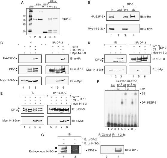
14-3-3ɛ binds to DP-3. (A) Coomassie stain showing bacterially expressed GST (track 4), GST-DP-3δ wt (track 5), GST-DP-3 5S (track 6) and 2 and 5 μg BSA (track 2 and 3, respectively) as protein standards. Molecular weight markers are shown in track 1. (B) COS7 cells were transiently transfected with expression vectors encoding myc-14-3-3ɛ (50 μg) and HA-E2F-5 (50 μg). The cells were harvested and used in a binding assay with either 5 μg of GST (track 2), GST-DP-3δ wt (track 3) or GST-DP-3 5S (track 4). The input levels of E2F-5 and 14-3-3ɛ are shown in track 1. (C) COS7 cells were transiently transfected with expression vectors encoding HA-E2F-5 (20 μg), DP-3δ wt (20 μg), myc-14-3-3ɛ (40 μg) and β-gal (5 μg) as an internal control. Cells were harvested and subjected to immunoprecipitation with anti-DP-3 antibody and immunoblotted with either anti-HA, anti-DP-3 or anti-myc as indicated. The immunoglobulin heavy chain (*) obscures the upper DP-3 polypeptide in the IB anti-DP-3 treatment. (D, E) COS7 cells were transiently transfected with the following amounts of the indicated expression vectors encoding DP-3δ wt (20 μg), DP-3 5S mutant (20 μg), myc-14-3-3ɛ (40 μg) and β-gal (5 μg) as an internal control. Cells were harvested and subjected to immunoprecipitation with either anti-DP-3 antibody (D) or anti-14-3-3ɛ antibody (E) and immunoblotted with either anti-DP-3 or anti-myc as indicated. The immunoglobulin heavy chain (*) obscures the upper DP-3δ polypeptide in the IB anti-DP-3 treatment. (F) In vitro-translated luciferase (tracks 2 and 3), HA-E2F-5 and DP-3δ wt (tracks 4–6) or HA-E2F-5 and DP-3 5S (tracks 7–9) were assessed for DNA binding on the E2F site taken from the E2A promoter as described. Track 1 shows the probe alone; wt indicates the presence of nonradiolabelled wild-type E2F site (100-fold excess) and − and + indicate the absence or presence of anti-HA antibody. The DP-3/E2F-5 complex and the supershift (SS) that occurs upon the addition of anti-HA are indicated. (G) Human ML-1 cells were harvested and subjected to immunoprecipitation with either anti-14-3-3ɛ or a control antibody, and then immunoblotted with either anti-DP-2 or anti-14-3-3ɛ as indicated. Endogenous 14-3-3ɛ and DP-2 are shown in tracks 1 and 2. The symbol • indicates a nonspecific band in the input extracts recognised by the anti-14-3-3ɛ antibody. Note that human DP-2 is equivalent to murine DP-3.