
Figure 3.
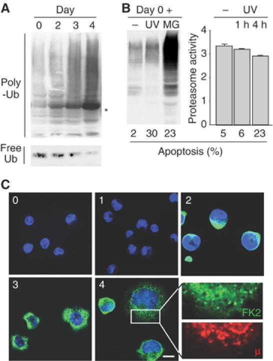
Accumulation of polyubiquitinated proteins in differentiating I.29μ+ cells. (A) Extracts of I.29μ+ cells induced with LPS for the indicated times were blotted with anti-Ub. The band indicated with an asterisk consists of μ-chains that crossreact with secondary antibodies. To detect free Ub, a denser gel was employed (bottom panel). (B) I.29μ+ cells were treated for 4 h with MG132 (MG) or exposed to ultraviolet light for 35 s and cultured for 4 h (UV, lane 6). In the right panel, proteasome activity was measured in unstimulated I.29μ+ cells cultured for 1 or 4 h after exposure to UV. The percentages of apoptotic cells are indicated at the bottom. (C) Immunofluorescent visualization of poly-Ub proteins during I.29μ+ differentiation. Cells were stained with Fk2 antibodies, recognizing poly-Ub proteins, and analyzed by confocal microscopy. Numbers indicate days after LPS stimulation. No overlapping between the Fk2 and anti-μ staining patterns was detected (see a particular at higher magnification in the bottom right panel). Bar: 5 μm.