
Figure 4.
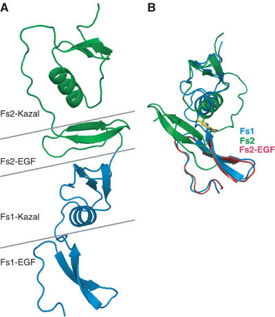
Structural flexibility in the Fs domains. (A) The structure of Fs12 fragment in the complex with Fs1 domain in blue and Fs2 in green. Horizontal lines mark the domain and subdomain boundaries highlighting the EGF subdomain of Fs2 sandwiched in between the two Kazal subdomains. (B) Superpositioning of Fs1 (blue) and Fs2 (green) domains using Kazal subdomains only, illustrating the EGF domains in opposite orientations. Disulphide bridge connecting the linker sequence from EGF subdomain to Kazal subdomain is shown for both Fs1 and Fs2. Similarity of the EGF domains is illustrated by the EGF domain of Fs2 alone in red, superimposed with equivalent part of Fs1 domain.